Publication

HealthCare

February 4, 2020

Dual Loading of Nanoparticles with Doxorubicin and Icotinib for the Synergistic Suppression of Non-Small Cell Lung Cancer

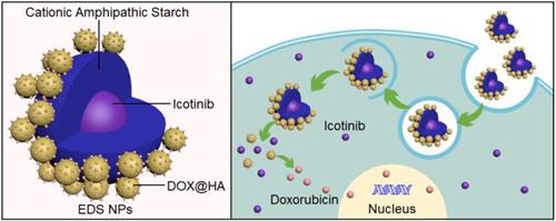
Background: Combination chemotherapy plays an important role in the clinical therapy of non-small cell lung cancer (NSCLC). However, the pharmacokinetic differences between drugs are an insurmountable barrier in traditional treatment. For the synergistic therapy of NSCLC, synergistic nanoparticles (EDS NPs) loaded with both an EGFR inhibitor and doxorubicin (DOX) were designed and prepared.

Methods: Erlotinib, apatinib and icotinib were evaluated for optimal combination with DOX in treatment of NSCLC via CCK-8 assay. Then the cationic amphipathic starch (CSaSt) and hyaluronic acid (HA) were applied to coencapsulate DOX and EGFR inhibitor to form the EDS NPs. EDS NPs were evaluated in NSCLC cell lines (A549, NCI-H1975 and PC9) and NSCLC xenograft mouse models.

Results: Icotinib was found to be the optimal synergistic drug in combination with DOX in the tested. Subsequently, icotinib and DOX were coencapsulated in the NPs. EDS NPs were roughly spherical with an average size of 65.7±6.2 nm and possessed stable loading and releasing properties. In the in vitro investigation, EDS NPs could efficiently deliver payloads into cells, exhibited cytotoxicity and produced strong anti-migration properties. In vivo hypotoxicity was confirmed by acute toxicity and hemolytic assays. The in vivo distribution showed that EDS NPs could enhance accumulation in tumors and decrease nonspecific accumulation in normal organs. EDS NPs significantly promoted the in vivo synergistic effects of icotinib and DOX in the mouse model.

Conclusions: The study suggests that EDS NPs possess noteworthy potential for development as therapeutics for NSCLC clinical chemotherapy.

Keywords: non-small cell lung cancer, combination chemotherapy, nanoparticles, icotinib, doxorubicin

Docetaxel and Doxorubicin Codelivery by Nanocarriers for Synergistic Treatment of Prostate Cancer

HealthCare

December 18, 2019

Docetaxel and Doxorubicin Codelivery by Nanocarriers for Synergistic Treatment of Prostate Cancer

Combination chemotherapy has been proven to be an efficient strategy for the treatment of prostate cancer (PCA). However, the pharmacokinetic distinction between the relevant drugs is an insurmountable barrier to the realization of their synergistic use against cancer. To overcome the disadvantages of combination chemotherapy in the treatment of PCA, targeted nanoparticles (NPs), which can codeliver docetaxel (DOC) and doxorubicin (DOX) at optimal synergistic proportions, have been designed. In this study, the DOC and DOX codelivery nanoparticles (DDC NPs) were constructed by hyaluronic acid (HA) and cationic amphipathic starch (CSaSt) through a self-assembly process. Human PCA cell lines (PC-3, DU-145, and LNCap) and mouse models were then used for evaluation in vitro and in vivo, respectively, of delivery and antitumor effects. The DDC NPs were spherical with rough surfaces, and the size and zeta potential were 68.4 ± 7.1 nm and -22.8 ± 2.2 mV, respectively. The encapsulation efficiencies of DOC and DOX in the NPs were 96.1 ± 2.3% and 91.4 ± 3.7%, respectively, while the total drug loading was 9.1 ± 1.7%. Moreover, the ratio of DOC to DOX in the DDC NPs was approximately 1:400, which aligned with the optimal synergistic proportions of the drugs. The DDC NPs exhibited excellent loading capacities, performed sustained and enzymatic release, and were stable in PBS, medium, and serum. After investigations in vitro, the DDC NPs were as effective as the dual drug combination in terms of cytotoxicity, antimigration, and apoptosis. Internalization results indicated that the DDC NPs could effectively deliver and fully release the payloads into PCA cells, and the process was mediated by the ligand-receptor interaction of HA with the CD44 protein. Low toxicity in vivo was confirmed by acute toxicity and hemolytic assays. The distribution in vivo showed that DDC NPs could enhance the accumulation of drugs in tumors and decrease nonspecific accumulation in normal organs. More importantly, DDC NPs significantly promoted the curative effect of the DOC and DOX combination in the PCA cell xenograft mouse model, indicating that the drugs with NPs did indeed act synergistically. This study suggests that the DDC NPs possess noteworthy potential as prospects for the development of PCA clinical chemotherapy.

https://www.frontiersin.org/journals/pharmacology/articles/10.3389/fphar.2019.01436/full

Sorafenib Inhibition of Hepatic Stellate Cell Proliferation in Tumor Microenvironment of Hepatocellular Carcinoma: A Study of the Sorafenib Mechanisms

July 16, 2014

Sorafenib Inhibition of Hepatic Stellate Cell Proliferation in Tumor Microenvironment of Hepatocellular Carcinoma: A Study of the Sorafenib Mechanisms

We investigated the mechanism and effects of sorafenib on hepatic stellate cell (HSC) viability and in the liver tumor microenvironment. The expression of α-smooth muscle actin (α-SMA) was measured immunocytochemically in the LX2 cells treated with differing concentrations of sorafenib. Changes in the platelet-derived growth factor (PDGF)-BB and tumor growth factor (TGF)-β1 concentrations were detected in the LX2 supernatant using an enzyme-linked immunosorbent assay (ELISA). Expressions of the extracellular signal-regulated kinase 1 (ERK1), ERK2, and Akt signaling pathways were measured using a western blot assay. The LX2 cells were cocultured with HepG2 cells for 24 h to observe their effects on HepG2 cell invasive ability.

(1) After treatment with various concentrations of sorafenib for 12, 24, 36, or 48 h, MTT assay showed that the viability of the treated LX2 cells was lower than in the controls.

(2) As sorafenib concentration and time of exposure increased, α-SMA expression became weaker in the treated cells.

(3) The PDGF-BB and TGF-β1 concentrations decreased with higher concentration, and longer exposures under the same sorafenib concentration.

(4) The ERK1, ERK2, and Akt expressions were identical between the treated and the control groups, but their phosphorylated expression decreased with increased concentrations of sorafenib.

(5) The invasive ability of the HepG2 cells induced by the LX2 gradually decreased as sorafenib concentrations increased. Sorafenib suppressed α-SMA expression, inhibited PDGF-dependent signaling pathways in HSCs, downregulated the PDGF-BB and TGF-β1 expression in the HSCs supernatant, and restrained viability of the HSCs, resulting in suppressed proliferation and invasion in the HepG2 cells.


https://pubmed.ncbi.nlm.nih.gov/24633454/

Current status of percutaneous vertebroplasty and percutaneous kyphoplasty – a review

HealthCare

October 7, 2013

Current status of percutaneous vertebroplasty and percutaneous kyphoplasty – a review

Percutaneous vertebroplasty (PV) and kyphoplasty (PK) are the 2vertebral augmentation procedures that have emerged as minimally invasive surgical options to treat painful vertebral compression fractures (VCF) during the last 2 decades. VCF may either be osteoporotic or tumor-associated. Two hundred million women are affected by osteoporosis globally. Vertebral fracture may result in acute pain around the fracture site, loss of vertebral height due to vertebral collapse, spinal instability, and kyphotic deformity. The main goal of the PV and PK procedures is to give immediate pain relief to patients and restore the vertebral height lost due to fracture. In percutaneous vertebroplasty, bone cement is injected through a minimal incision into the fractured site. Kyphoplasty involves insertion of a balloon into the fractured site, followed by inflation-deflation to create a cavity into which the filler material is injected, and the balloon is taken out prior to cement injection. This literature review presents a qualitative overview on the current status of vertebral augmentation procedures,especially PV and PK, and compares the efficacy and safety of these 2 procedures. The review consists of a brief history of the development of these 2 techniques, a discussion on the current research on the bone cement, clinical outcome of the 2 procedures, and it also sheds light on ongoing and future research to maximize the efficacy and safety of vertebral augmentation procedures.

Keywords: Bone Cements – therapeutic use, Fractures, Compression – surgery, Kyphoplasty – trends, Spinal Fractures – surgery, Surgical Procedures, Minimally Invasive – trends, Vertebroplasty – trends

Advances in diagnosis and treatment of hilar cholangiocarcinoma – A review

HealthCare

August 1, 2013

Advances in diagnosis and treatment of hilar cholangiocarcinoma – A review

Hilar cholangiocarcinoma (HC) is a rare tumor that causes devastating disease. In the late stages, this carcinoma primarily invades the portal vein and metastasizes to the hepatic lobes; it is associated with a poor prognosis. HC is diagnosed by its clinical manifestation and results of imaging techniques such as ultrasound, computed tomography, magnetic resonance cholangiopancreatography, endoscopic retrograde cholangiography, and percutaneous transhepatic cholangiography. Preoperative hepatic bile drainage can improve symptoms associated with insufficient liver and kidney function, coagulopathy, and jaundice. Surgical margin-negative (R0) resection, including major liver resection, is the most effective and potentially curative treatment for HC. If the tumor is not resected, then liver transplantation with adjuvant management can improve survival. We conducted a systematic review of developments in imaging studies and major surgical hepatectomy for HC with positive outcomes regarding quality of life.

Kyphoplasty for the Treatment of Pain Distant to Osteoporotic Thoracolumbar Compressive Fractures

August 1, 2013

Kyphoplasty for the Treatment of Pain Distant to Osteoporotic Thoracolumbar Compressive Fractures

Vertebral fractures are one of the most common osteoporotic fractures. We sought to investigate the incidence of distant pain after osteoporotic vertebral compressive fracture (OVCF) at the thoracolumbar junction, and to explore the effect of kyphoplasty in the treatment of distant pain post-OVCF. Eighty-seven patients diagnosed OVCF between T11 and L2 were included in the study. The region of pain and its proximity to the thoracolumbar compressive fracture was recorded. For pain management, all patients received kyphoplasty. The follow-up period was every 3 months for 1-year post-surgery. The Visual Analogue Scale (VAS) and Oswestry Disability Index (ODI) were used pre-operatively, post-operatively, and at 3-, 6-, and 12-month follow-ups to assess patient status. All patients completed the operation, with 72 patients having focal pain over the compression fracture. Eleven cases also had pain distal to the fracture region in the following areas: lower back, near the iliac crest (n = 6), the groin (n = 3), and the trochanteric region (n = 2). Four cases had pain in distant to the fracture: lower back, near iliac crest (n = 3), and the trochanteric region (n = 1). All patients had a significant improvement in clinical symptoms. The average VAS and the ODI decreased significantly pre-operatively to post-operatively (p < 0.05). In addition to focal tenderness, many patients with thoracolumbar compression fractures may have pain distant to the fracture. This can be successfully treated using kyphoplasty. This phenomenon also indicates that patients at risk of osteoporosis who also have lower back pain should not neglect the potential for a thoracolumbar fracture.

Resveratrol Suppresses Microcirculatory Disturbance in a Rat Model of Severe Acute Pancreatitis

HealthCare

April 2, 2013

Resveratrol Suppresses Microcirculatory Disturbance in a Rat Model of Severe Acute Pancreatitis

The present study sought to understand the mechanisms of attenuation of severe acute pancreatitis (SAP) by resveratrol (RES). SAP was experimentally induced in rats by injection of 4 % sodium taurocholate in the retrograde pancreatic duct. Three study groups were evaluated: Group I (sham-operated animals), Group II (SAP animals), and Group III (SAP animals treated with RES at 20 mg/kg/body weight, 5 min after induction of SAP). The study outcomes were histopathologic changes and alterations in biochemical markers: plasma renin activity and levels of angiotensin II, endothelin, and nitric oxide in plasma. Biochemical markers were evaluated at 3, 6, and 12 h after induction of SAP. SAP was associated with significant (p < 0.05) histopathologic changes (saponification spots in the intraperitoneal cavity, severe pancreatic edema, blood congestion, varying degrees of necrosis, etc.), as well as with elevation of biochemical markers in blood plasma. RES treatment significantly (p < 0.05) attenuated changes of both histopathologic and biochemical markers induced by SAP. In conclusion, this study provides evidence that RES treatment is a promising therapeutic approach to suppress microcirculatory disturbance in SAP.